IMAGING
Radiographs (X-rays)
Imaging that helps evaluate bones, chest structures, abdominal patterns, heart silhouette and other anatomy when there is a relevant clinical question.
THE SHORT VERSION
Testing helps answer a clinical question.
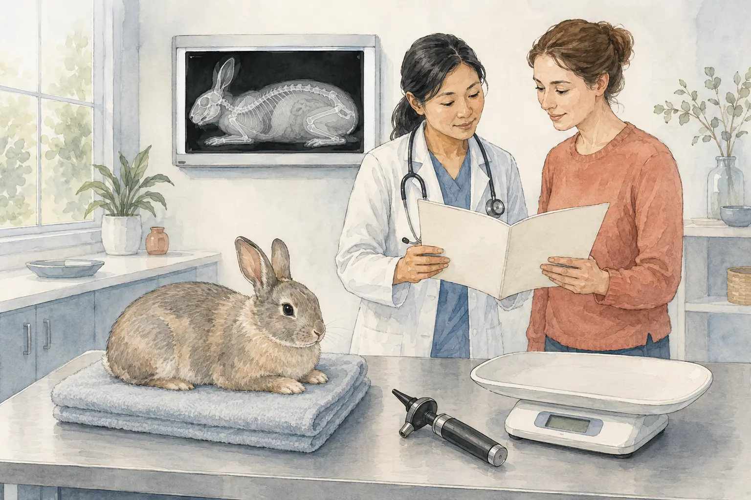
Radiographs use controlled X-ray exposure to create images of internal structures. Chest, abdominal and orthopedic views answer different questions, and more than one positioning view may be needed.
What is this test?
Radiographs use controlled X-ray exposure to create images of internal structures. Chest, abdominal and orthopedic views answer different questions, and more than one positioning view may be needed.
What information can it provide?
- Chest radiographs can help evaluate lung patterns, heart silhouette, airways and fluid-related concerns.
- Abdominal radiographs can assess organ outlines, intestinal gas, some foreign material, urinary stones and other structural patterns.
- Orthopedic radiographs help examine fractures, arthritis, joint alignment and other skeletal concerns.
- Appropriate positioning, several views and professional interpretation influence diagnostic value.
When might your veterinarian recommend it?
- Investigate cough, breathing changes, limping, trauma, abdominal pain or suspected obstruction.
- Compare known disease progression or investigate a specific physical-examination finding.
- Support an individualized baseline discussion when the veterinarian believes the information could change care.
What can owners do before the visit?
- Ask whether fasting, sedation or careful handling is recommended.
- Share prior imaging so the veterinarian can compare changes.
- Tell the clinic about pain, breathing difficulty, pregnancy or prior anesthesia concerns.
What can this test not tell you by itself?
- Radiographs cannot identify every early disease or characterize all soft tissues.
- One lateral image is not equivalent to a complete diagnostic study.
- Not a universal screening recommendation: routine baseline radiographs of healthy animals are practice- and patient-dependent, not an automatic fixed-schedule requirement.
Do not change medication, interpret an abnormal number as a diagnosis or delay urgent treatment without direction from your veterinary team.
Useful questions for your appointment
- What specific clinical question are these images intended to answer?
- How many views are necessary, and would ultrasound or another test add information?
- Is sedation recommended for safety, comfort or more accurate positioning?